
ImekaTM: The gold standard of clinical white matter imaging
Our technology is powerful and non-invasive
Imaging means so much more with powerful processing tools
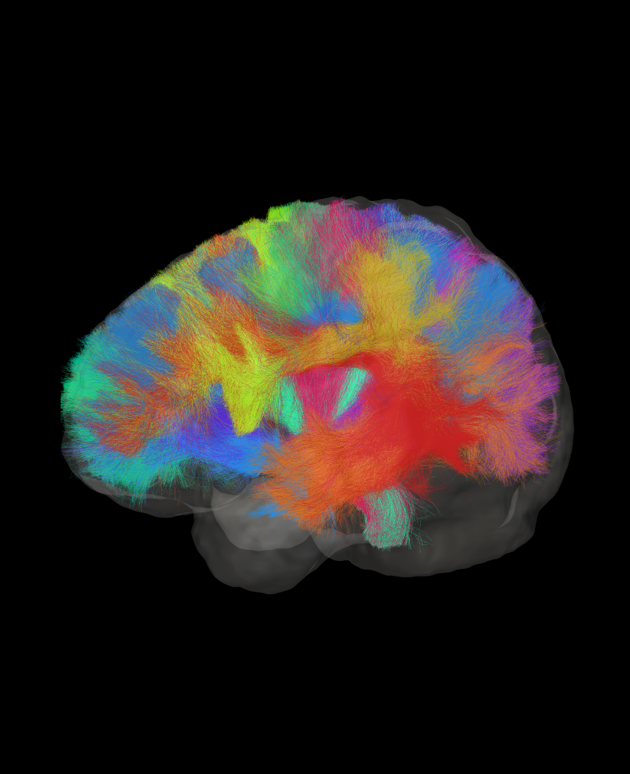
Advanced white matterA large network of nerve fibers that communicate between different areas of the brain. imaging is a readily available technology that picks up microstructural properties of the central nervous systemResponsible for receiving, processing, and responding to sensory information in the body.. Data can be collected in as little as five minutes on a standard MRIMagnetic Resonance Imaging is a medical imaging technique used in radiology to form pictures of the anatomy and the physiological processes inside the body. machine.
ANDI’s focus is on greater detail. We do so by combining white matterA large network of nerve fibers that communicate between different areas of the brain. imaging with AIArtificial Intelligence to better analyze white matterA large network of nerve fibers that communicate between different areas of the brain. MRIMagnetic Resonance Imaging is a medical imaging technique used in radiology to form pictures of the anatomy and the physiological processes inside the body. signals. Want to see how ANDIAdvanced Neuro Diagnostic Imaging can benefit you and your practice?
Learn more
Meet the people that make ImekaTM
Meet our executives
Louis-Philippe Bourgault
TBD
Jean-Christophe Houde
Jean-Christophe has a bachelor’s and a master’s degree in computer science with a specialization in image analysis. He is in charge of managing the operations unit at Imeka and ensures Imeka stays at the forefront of the field by keeping up to date on developments in the field of medical imaging analysis. His in-depth knowledge of dMRI and hands-on experience come from his role as a research associate at the Sherbrooke Connectivity Imaging Lab (SCIL), which is headed by Maxime Descoteaux (Imeka’s co-founder and chief science officer).
Meet our advisors


Maxime Descoteaux
Maxime received a PhD from Inria Sophia-Antipolis and did a postdoctoral fellowship in computer science and neuroimaging at NeuroSpin, CEA Saclay in Paris. He is a world-renowned expert in advanced white matterA large network of nerve fibers that communicate between different areas of the brain. imaging and medical imaging. Maxime supervises the science, research, and software developments at Imeka.
In addition to his work with Imeka, Maxime leads the Sherbrooke Connectivity Imaging Lab (SCIL), known for its integrated approach to advanced white matterA large network of nerve fibers that communicate between different areas of the brain. imaging. Maxime’s credentials include:
- 16,523 citations; 125+ journal papers; h-index 61, i10-index 186
- Member of the Royal Society of Canada’s College of New Scholars, Artists, and Scientists
- Full professor, Computer Science, University of Sherbrooke, QC, Canada
- Leads a lab of 15 members (MSc, PhD, postdocs)
- International leader and conference/workshop organizer in diffusion MRIMagnetic Resonance Imaging is a medical imaging technique used in radiology to form pictures of the anatomy and the physiological processes inside the body.
Pierre-Marc Jodoin
Pierre-Marc is a world expert in computer vision and deep learning. He received a PhD in computer vision from the University of Montreal, followed by a postdoc at Boston University. He is currently a full professor at the University of Sherbrooke, working on the development of deep learning solutions to various digital imaging problems. Pierre-Marc supervises software development at Imeka.
In addition to his work with Imeka, Pierre-Marc runs the Videos & Images Theory and Analytics Laboratory (VITAL), which is 100% focused on deep learning–based techniques. VITAL focuses on researching state-of-the-art algorithms in image processing, computer vision, video analytics, medical imaging, and statistical models.
Meet the whole team
Matthieu Dumont
Applied Integrator in Diffusion Imaging
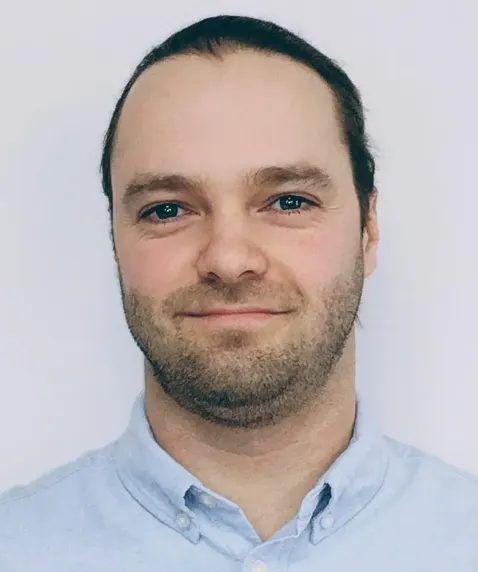
Francis Houde
Clinical Science Liaison
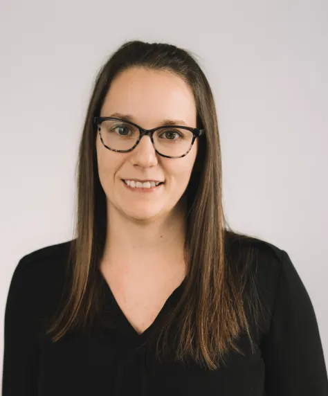
Loïka Maltais
Project Manager and Software Quality Assurance
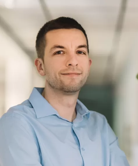
Nil Goyette
Software Developer
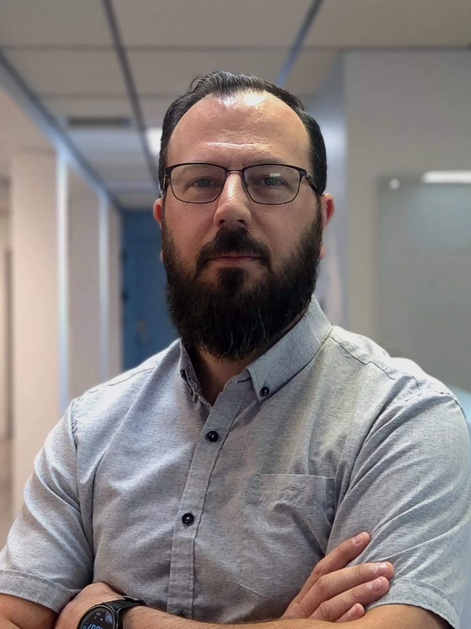
Jean-Sébastien Frerot
System Administrator
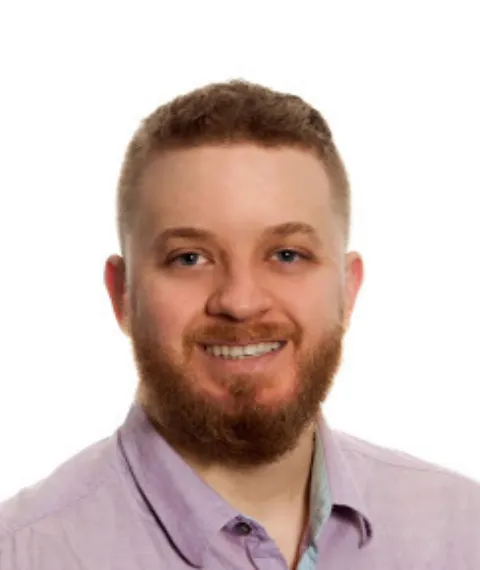
Chris Majcher
Software Developer in Medical Imaging
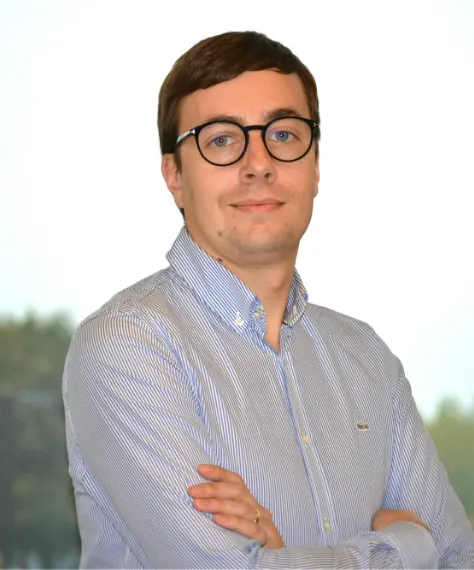
Jean-Sébastien Lord
Quality Manager
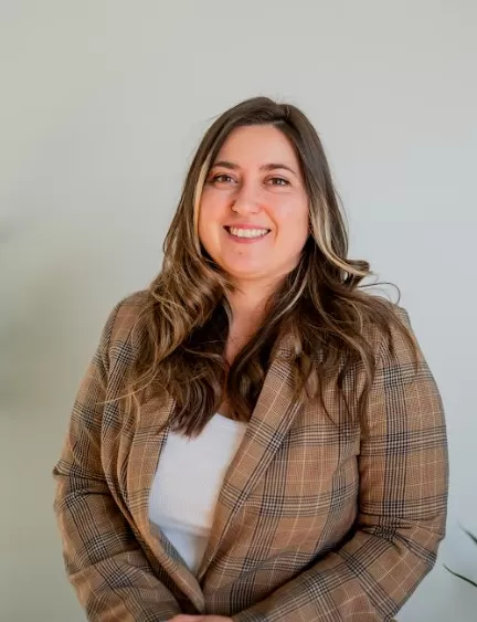
Amélia Bourbonnais
Administrative Coordinator & Customer Support
Join us!
Let’s work together!
Apply now