We are building the future of brain imaging
At ImekaTM, we put patients first. That’s why we are committed to enhancing our clinical tools.
What we aim to measure
Imeka’s pipeline
What we aim
to measure
Measures key markers of neuropathology.
Measures therapeutic effects.
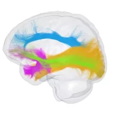
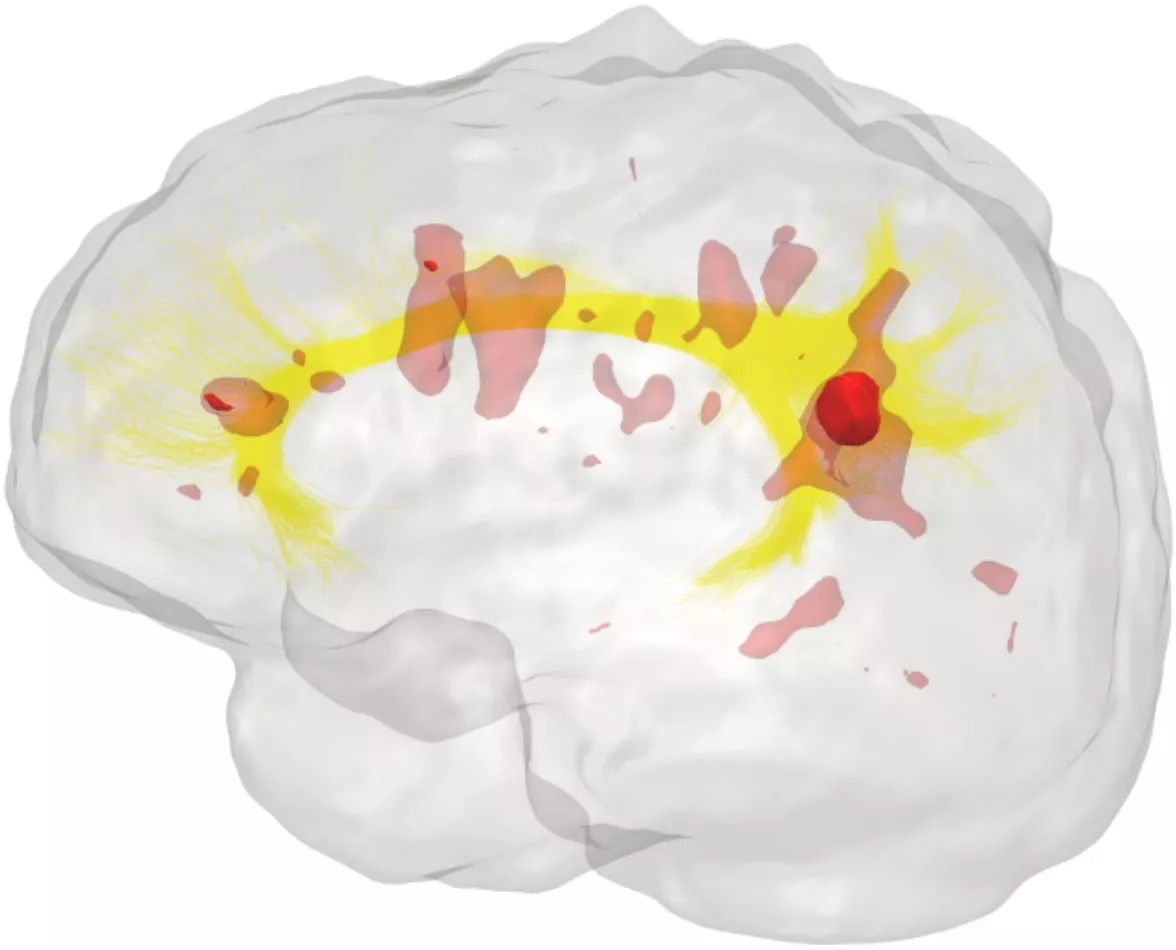
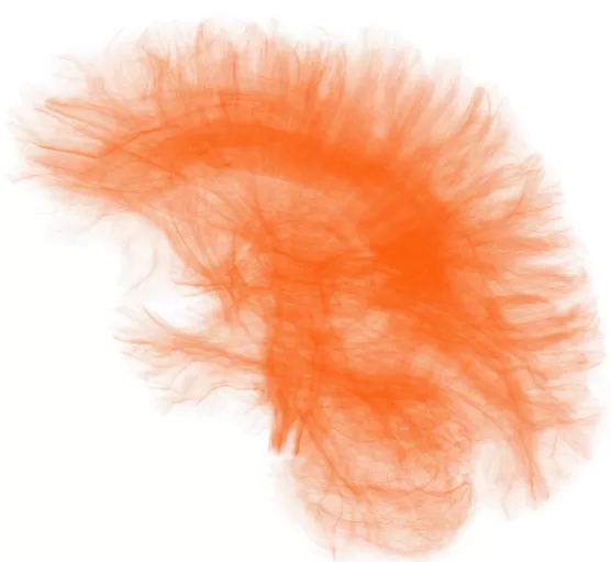
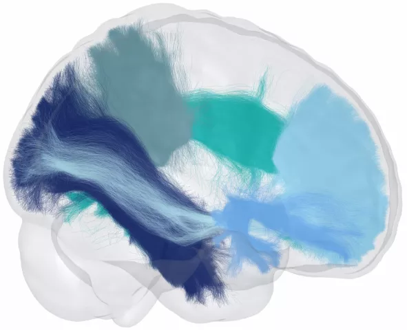
Provides virtual dissections of 50+ fiber bundles—robust to crossing fibersComplex geometrical configurations that present a challenge for diffusion MRI reconstruction and fiber tracking..
The ultimate goal of our white matterA large network of nerve fibers that communicate between different areas of the brain. imaging technology is to find non-invasive markers that are sensitive to pathology and can quantify damage and/or therapeutic changes.
Imeka’s pipeline
Imagine a future where you can determine the exact rehabilitation protocol needed to get your patients back to normal.
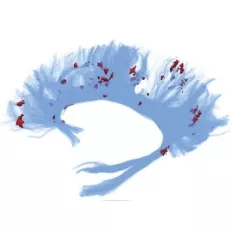
We identify and quantify neuroinflammationInflammation of the nervous tissue in the central nervous system. with a high degree of sensitivity and specificity. This is performed by measuring the free water volumeAn analytical method for diffusion MRI that models extracellular free water and water that is in the vicinity. found in a patient’s white matterA large network of nerve fibers that communicate between different areas of the brain..
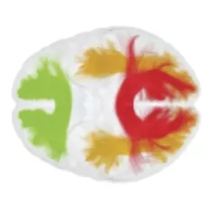
We identify myelin integrityThe integrity of myelin. Myelin is an insulating layer (or sheath) that forms around nerves. by the location, extent, and degree of demyelinationA condition that causes damage to the protective covering (myelin sheaths) of nerve fibers. and remyelinationA phenomenon by which new protective coverings (myelin sheaths) are generated around axons. found in a patient’s brain. Our clinical software can even identify pathology in normal-appearing white matterA large network of nerve fibers that communicate between different areas of the brain..
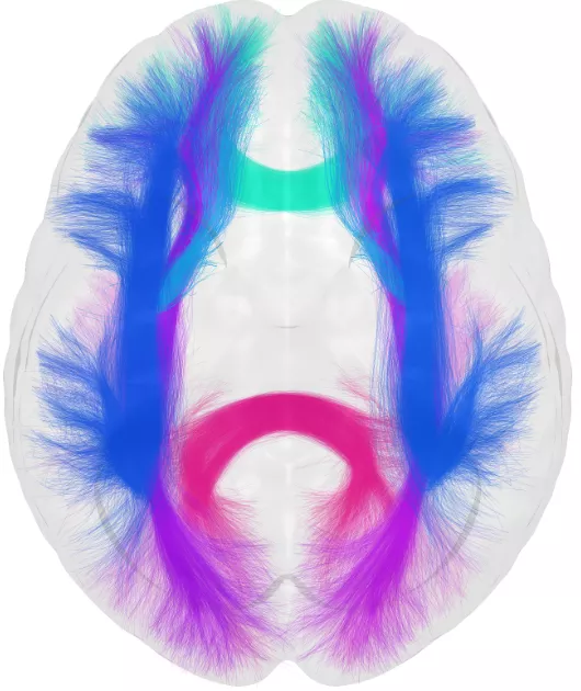
We identify, measure, and characterize axonal integrityThe integrity of axons. Axons are long cellular projections of neurons that relay electrical and biochemical signals in nerves and white-matter tracts. and white matterA large network of nerve fibers that communicate between different areas of the brain. health with a high degree of precision through free water movementA measurement derived from DTI that assesses the presence of extracellular fluid in brain tissue..
We are robust to crossing fibersComplex geometrical configurations that present a challenge for diffusion MRI reconstruction and fiber tracking.. We go beyond diffusion tensor imaging trackingAn MRI technique that visualizes white matter tracts., which assumes single-directional tracts.
Let’s work together!
Contact us